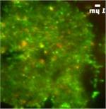
In this movie, localization of GFP-tagged myo1e (green,) and RFP-tagged clathrin (red) at the site of endocytosis was examined using TIRF microscopy.
Experimental Methods
These are some of the experimental techniques commonly used in the lab:
- Live cell imaging and analysis of dynamics of fluorescently tagged proteins.
- Physiological characterization of renal functions in mice.
- Mouse model of type I diabetes (streptozotocine-induced diabetes).
- Histology, immunohistochemistry, and electron microscopic analysis of animal tissue samples.
- Measurements of cell migration rates using wound-healing model and Boyden chamber migration assays.
- Use of adenoviral vectors for recombinant protein expression and protein knockdown.
- Protein-protein interaction assays (pulldowns, immunoprecipitation, yeast two-hybrid).
This time-lapse movie of wound healing was collected after scraping a monolayer of epithelial cells with a needle and allowing cells to migrate into the wound.